Home

Revocation Simplify equilibrium measuring lvot diameter Chip Typically Candy

The LVOT diameter was obtained from LVOT images in the long-axis view.... |  Download Scientific Diagram
The LVOT diameter was obtained from LVOT images in the long-axis view.... | Download Scientific Diagram

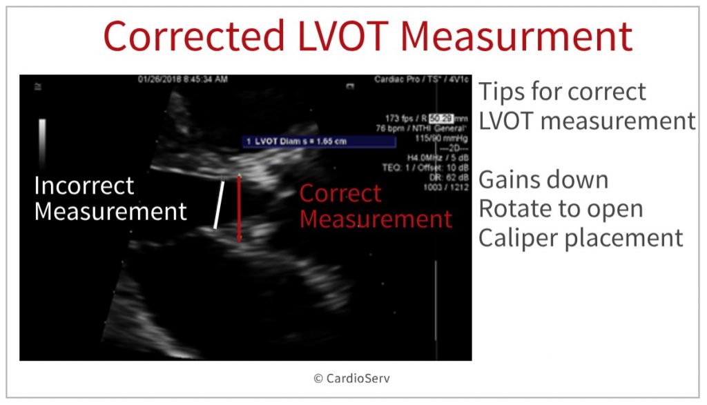
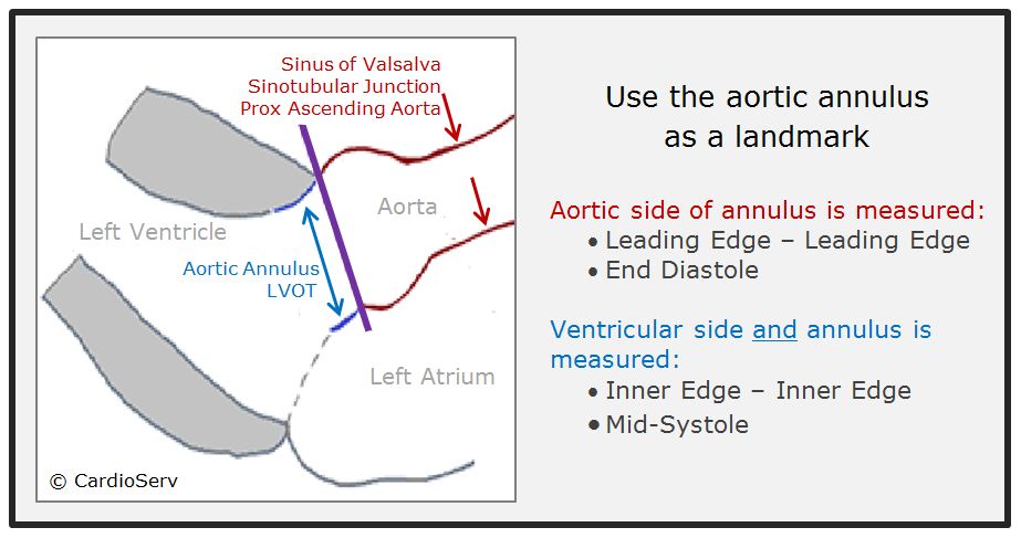
How to Master Aortic Measurements With These 5 Techniques Cardioserv

Aortic valve area calculation in aortic stenosis by CT and Doppler  echocardiography. | Semantic Scholar
Aortic valve area calculation in aortic stenosis by CT and Doppler echocardiography. | Semantic Scholar

How to Master Aortic Measurements With These 5 Techniques Cardioserv

ECHOCARDIOGRAPHY: How to measure the LVOT? - YouTube

Impact of anatomical variations of the left ventricular outflow tract on  stroke volume calculation by Doppler echocardiography in aortic stenosis -  Pu - 2020 - Echocardiography - Wiley Online Library
Impact of anatomical variations of the left ventricular outflow tract on stroke volume calculation by Doppler echocardiography in aortic stenosis - Pu - 2020 - Echocardiography - Wiley Online Library

Optimal measurement of LVOT diameter is shown on TTE (a) and TEE (b).... |  Download Scientific Diagram
Optimal measurement of LVOT diameter is shown on TTE (a) and TEE (b).... | Download Scientific Diagram

Accurate stroke volume (SV) estimation: SV = LVOT area × LVOT VTI. a... |  Download Scientific Diagram
Accurate stroke volume (SV) estimation: SV = LVOT area × LVOT VTI. a... | Download Scientific Diagram

Aorta and aortic valve - basic principle of 2D echo assessment

Effect of assessing velocity time integral at different locations across  ventricular outflow tracts when calculating cardiac output in neonates |  European Journal of Pediatrics
Effect of assessing velocity time integral at different locations across ventricular outflow tracts when calculating cardiac output in neonates | European Journal of Pediatrics

Left Ventricular Outflow Tract: Intraoperative Measurement and Changes  Caused by Mitral Valve Surgery | Thoracic Key
Left Ventricular Outflow Tract: Intraoperative Measurement and Changes Caused by Mitral Valve Surgery | Thoracic Key
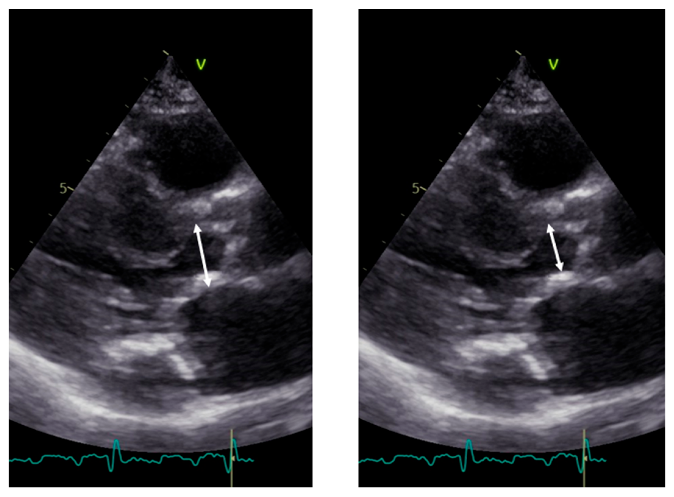
Aortic Regurgitation | Radiology Key

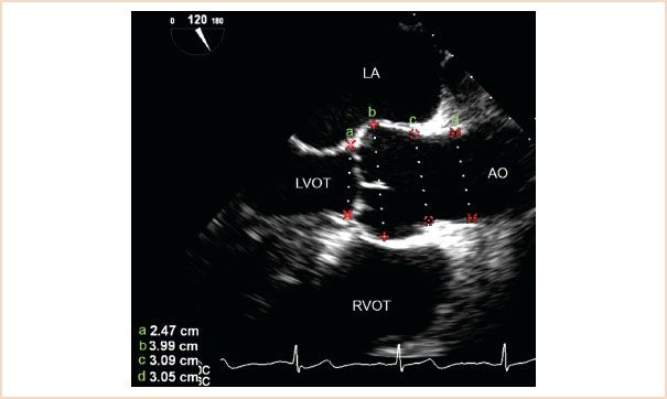
Diagnostics | Free Full-Text | Pitfalls and Tips in the Assessment of  Aortic Stenosis by Transthoracic Echocardiography
Diagnostics | Free Full-Text | Pitfalls and Tips in the Assessment of Aortic Stenosis by Transthoracic Echocardiography
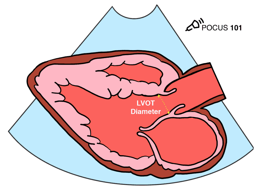
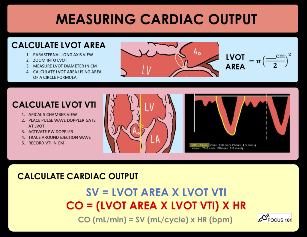
Measuring Cardiac Output with Echocardiography Made Easy - POCUS 101

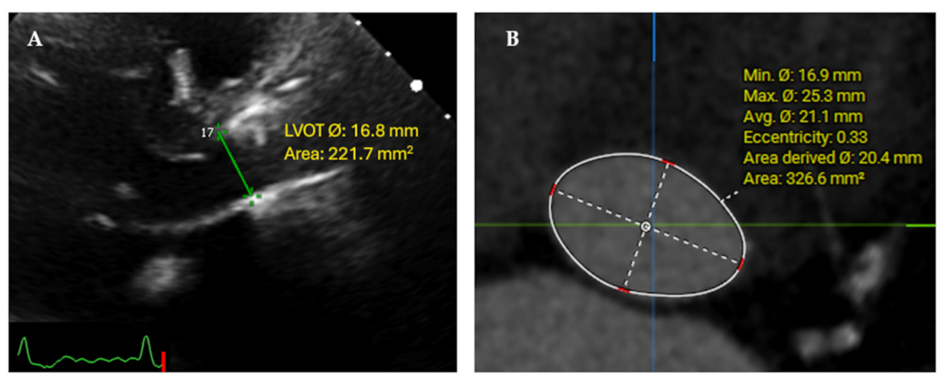
JCM | Free Full-Text | Core Lab Adjudication of the ACURATE neo2  Hemodynamic Performance Using Computed-Tomography-Corrected Left  Ventricular Outflow Tract Area
JCM | Free Full-Text | Core Lab Adjudication of the ACURATE neo2 Hemodynamic Performance Using Computed-Tomography-Corrected Left Ventricular Outflow Tract Area

FINALLY: How to Correctly Measure the Aorta!

Measuring Cardiac Output with Echocardiography Made Easy - POCUS 101

Accurate Measurement of Left Ventricular Outflow Tract Diameter: Comment on  the Updated Recommendations for the Echocardiographi
Accurate Measurement of Left Ventricular Outflow Tract Diameter: Comment on the Updated Recommendations for the Echocardiographi

PLAX view optimized for measuring the LVOT diameter. | Open-i

Imaging of Low-Gradient Severe Aortic Stenosis - ScienceDirect

kazi ferdous on X: "-Aortic annulus and LVOT diameter are measured in mid  systole. - Ascending aorta in end diastole -Mitral valve area, mitral  annulus, tricuspid annulus are measured in early or
kazi ferdous on X: "-Aortic annulus and LVOT diameter are measured in mid systole. - Ascending aorta in end diastole -Mitral valve area, mitral annulus, tricuspid annulus are measured in early or

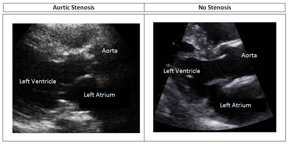
Aortic Stenosis and Mismatch Values Cardioserv

Measuring Cardiac Output with Echocardiography Made Easy - POCUS 101

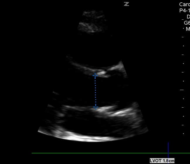
LVOT measurement 1. PLX view zoom mode 2. Measure ... | GrepMed